
Every year, tens of thousands women are diagnosed with ovarian tumors. These can range from benign to varying degrees of malignancy. A correct diagnosis is important to determine the most appropriate treatment.
New strategy for classification
The international research group IOTA, coordinated from Leuven, developed a new "two-step" strategy to better classify ovarian tumors. In the first step, they look at some features that can already classify many of the cysts without the need for a computer. For the ovarian tumors where this is not possible, a next step follows based on a statistical model (ADNEX) that uses ultrasound images to determine the likelihood that the tumor is benign or a certain type of malignant tumor.
Recently, a team of researchers from KU/UZ Leuven, Imperial College London (United Kingdom) and the University of Lund (Sweden), published the results of a large international study in which they validated the IOTA strategy in 4,905 patients in 17 international centers. They showed that this strategy can well indicate which patients belong to a higher risk group, and this is fully consistent with the O-RADS system, developed in 2020 by the American College of Radiology and the IOTA researchers at Leuven.
Dr. Stefan Timmerman, gynaecologist in training and first author of the study said, "Our strategy proved very successful in correctly recognizing benign neoplasms. Furthermore, the model was also able to distinguish between borderline (between benign and malignant), stage I cancer, stage II-IV cancer or metastatic tumors."
Importance for follow-up
A correct diagnosis helps the physician determine whether or not surgery is appropriate.
Prof. Dr. Wouter Froyman, gynecologist at University Hospitals Leuven and principal investigator of the study, says: "The added value of our strategy is that it offers a more refined diagnosis of potentially malignant tumors, which increases the patient's chances of survival. And if it turns out to be a benign tumor, it often requires no or only minimally invasive surgery, which saves patients a great deal of inconvenience and is also cost-effective for the health care system. This is an important step forward toward the most optimal care for patients with ovarian tumors."
This is an important step forward toward the most optimal care for patients with ovarian tumors.Prof. Dr. Wouter Froyman, gynecologist at UZ Leuven
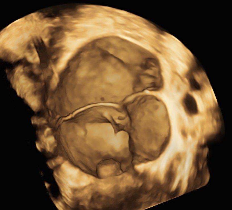
The model uses visual characteristics of the tumour, as seen in this ultrasound 3D image, to determine a risk score. This example depicts is a benign ovarian tumour.