Giant cell arteritis
Giant cell arteritis is the most common type of vasculitis, a inflammatory disease of the arteries. This disorder is also called arteritis temporalis, and occurs mostly in people over 50. About 1% of all women and 0.5% of all men come across it in their lives. GCA affects the larger arteries: the aorta or main artery and its branches. The cause is unknown, but typical symptoms are headaches, fatigue, stiffness in the shoulder and pelvic belt and pain in the jaws when chewing.
Giant cell arteritis can be detected with the help a PET scan, during which the radioactive substance fluorodeoxy-glucose (FDG) is administered. This substance settles in those places in the body where glucose is absorbed the most, indicating inflammation. In the case of GCA this is usually in the wall of the large coronary artery, the blood vessels going to the arms, and some blood vessels in the head.
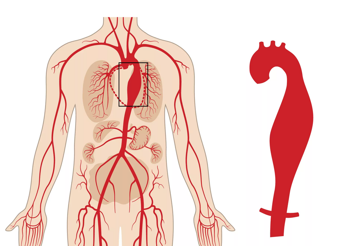
Even though treatment with cortisone can largely cure the disorder, there are a number of risks connected to GCA. One of these is the widening of the diameter of the aorta, with Eén daarvan is het verbreden van de diameter van de aorta, resulting in possible rupture (an aortic aneurysm). This type of complication is rare and often doesn't occur until after a couple of years, but until now it was not known which patients were most at risk of such risky widening.
Close monitoring of high-risk patients
A prospective study, performed at the UZ Leuven department of internal medicine, now shows that patients whose blood vessels absorbed the most FDG during the PET scan - indicating more inflammation - also showed the most widening of the aorta afterwards. For the study, at total of 106 GCA patients were monitored for 12 years. At diagnosis, they all underwent an FDG-PET scan. After that patients returne annually for a CT scan of the aorta, during which the diameter of the aorta was accurately measured at six different heights.
If patients clearly have an inflamed aorta, it is recommended that they are closely monitored in order to recognise an impending aortic aneurysm in time. Prof. dr. Daniel Blockmans
Prof. dr. Daniel Blockmans, doctor-specialist general internal medicine at UZ Leuven: “The PET-scan can play an important role while assessing the further course of giant cell arteritis. The study shows that it is recommended that patients with the condition should undergo a PET scan at diagnosis. If they a clearly inflamed aorta, they can monitored closely, in order to recognise an impending aortic aneurysm in time.”
Want to find out more?
- The study, exclusively performed at UZ Leuven, was published in the medical journal Annals of Internal Medicine.
- The study was done as part of Dr Lien Moreel's PhD research. Dr Moreel works partly as an ASO at the department of general internal medicine at UZ Leuven. For her research, she conducted several retrospective and prospective observational clinical studies on the diagnosis, clinical patterns and follow-up of polymyalgia rheumatica and giant cell arteritis.
At UZ Leuven most GCA patients undergo a PET scan as a standard.