- Collaboration with the cardiology department (electrophysiology)
- During the procedure, a 3D image of the ventricle that is being treated is generated. Based on artificial intelligence, the anatomy is automatically extracted from these images. In addition, a planning of the treatment is also automatically generated (video). After the verification by the specialist, the planning can be shown together with the interventional radioscopic images. This way the treatment can be executed very precisely.
AI based anatomy recognition: in this example the anatomy of the left atrium is automatically detected in 3D medical images via artificial intelligence. This method also detects the various sub-regions of the atrium.
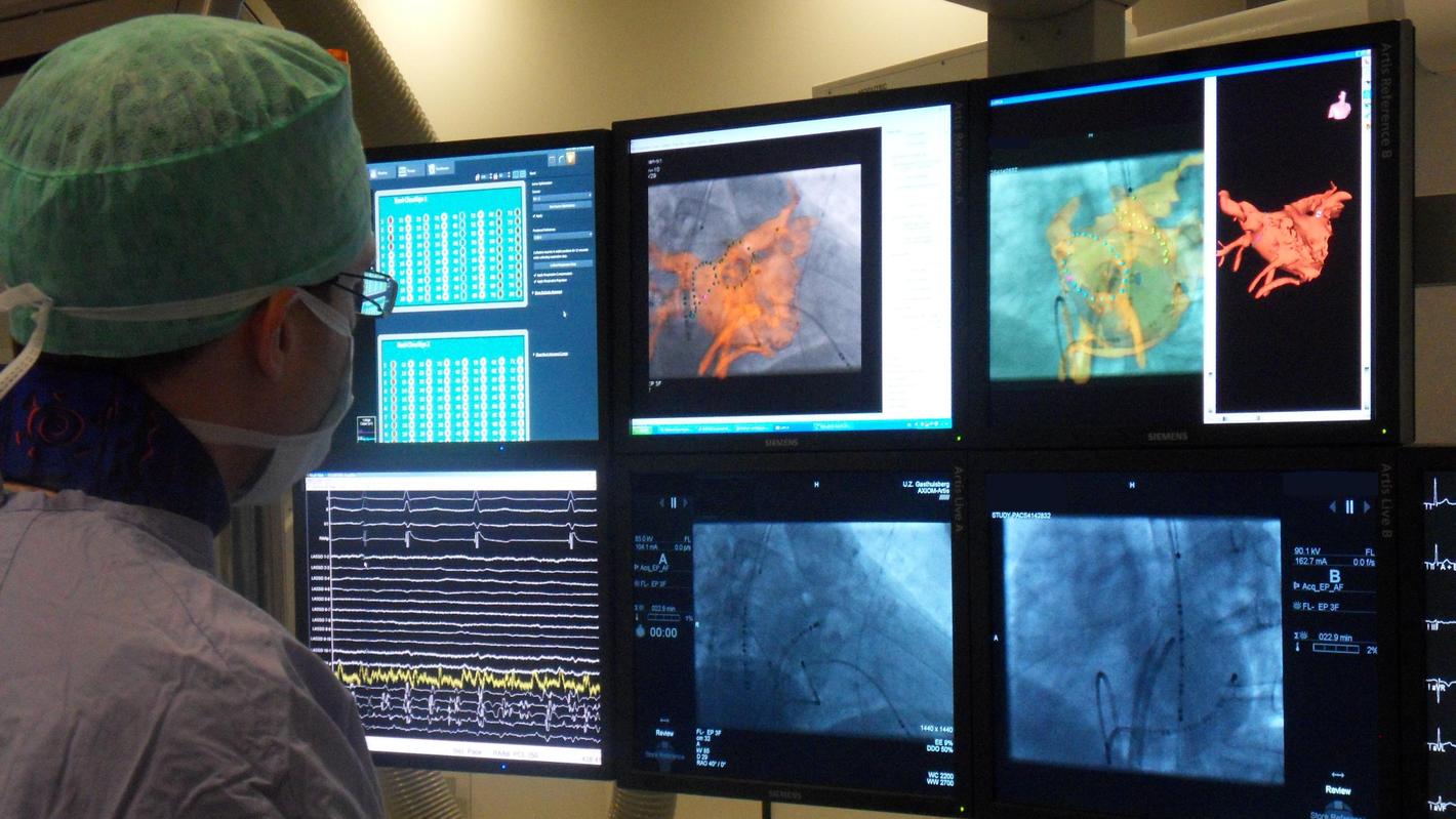
The cardiologist can use 3D imaging and the automatically detected anatomy and planning to treat cardiac arrhythmias via catheters. The use of 3D information allows for a much more precise execution of the treatment.