Diagnosing Alzheimer's today
Alzheimer's Disease, one of the most common causes of dementia, is a big problem for our ageing population. Today in Belgium more than 220,000 people suffer from the disease, and together with them thousands of relatives and friends that have to watch their loved ones deteriorate. The World Health Organisation has called Alzheimer's, in addition to cancer and diabeted the most expensive diseas of our time.
There is no miracle cure for Alzheimer's. The last couple of years various candidate-medicines have been discarded because of disappointing results in the last stage of the clinical trials. Experts believe that existing medication would be more effective if it could be administered in a much earlier stage, before the brain is affect and before symptoms such as memory loss or speech problems occur.
Expensive brain scans are not an option for everyone. But a simple check-up at the ophthalmologist could be.
But could you screen risk patients - or even the entire population - for Alzheimer's? Expensive brain scans are not an option. But a simple check up at the ophthalmologist could be.
The eye as the window to the brain
“In 2015 researchers from Minnesota (USA) published exceptional results where they were able to detect accumulations of the amyloid beta protein on the retina of mice that developed Alzheimer's, even before they had any symptoms,” prof. dr. Ingeborg Stalmans, ophthalmologist at UZ Leuven, explains. “These results were very controversial at the time, and a lot of researchers did not put much stock in it. Even though it is not that strange to study the eye and find out more about the development of chronic diseases. You only have to think about the study of veins in the eye to detect hypertension or the risk of a heart attack. The big advantage is of course the fact that the eye is transparant and allows for a very simple and non-invasive view into the body and the brain.”
A multidisciplinary team
Professor Stalmans and her team, dr. Karel Van Keer and dr. ir. Sophie Lemmens, have decided to explore this further and have joined forces with biologists and engineers. The team of professor Lieve Moons and postdoctoral researcher Lies De Groef of the animal physiology and neurobiology department at the KU Leuven will take care of the basic research and study the formation of protein accumulations in the retina of mice, for various brain disorders. “We are looking for abnormal protein accumulations that are typical for diseases such as Alzheimer's, Parkinson's, Huntington's etc.”, they explaint. “We hope to find a 'finger print' for every disease that can lead to a better diagnosis.”
For the development of the required camera technology, UZ Leuven and KU Leuven went to its 'neighbour': imec. “For more than ten years we have been working with hyperspectral camera's and we are making them as small, fast and user-friendly as possible camera’s”, says Andy Lambrechts, programme director for Hyperspectral Technology at imec. “Whereas a normal camera can only detect three colours or frequencies (red, blue and green), a hyperspectral camera can divide the light captured into tens or hundreds of frequencies. Given the fact that every substance, in this case protein, will reflect specific frequencies to the camera, it is possible to identify them. We call this a spectral finger print that is specific for certain type of protein.”
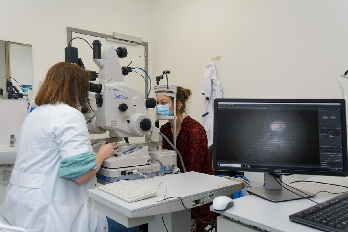
To make the bridge between imec's hyperspectral camera and the actual application needed by the biologists and the ophthalmologists, a lot of team work was required. “We integrated the camera in a microscope for the examination of tissue samples, and into a fundus camera for eye examinations in patients,” says dr. ir. Sophie Lemmens, who has devoted her doctoral research on this.
The team got the help of VITO. Professor Patrick De Boever, former project manager at the VITO Health Unit: “For the ophthalmologists it is especially important to optimise the speed of the camera, as it has to make images in-between the patient's eye blinking.” Ir. Jan Theunis, current project manager at VITO Health, adds: “Based on the hyperspectral images, we subsequently developed smart algorithms in order to recognize an Alzheimer's pattern. These algorithms can then be used to detect the disease with a simple eye test.”
I really believe that if we join forces, we can fight this disease and make the life of all those millions of patients more bearable.Prof. dr. Ingeborg Stalmans, ophthalmologist at UZ Leuven
Promising and for the near future
Normally this type of research does not pay off that quickly in practice, but this could happen pretty fast. The equipment is available and does not require a lot of extra development. In additions, the test is safe for patients, because it only uses visible light, not potentially dangerous laser light.
“It is now important to show how early in the development of the disease we detect protein plaques in the eye”, prof. dr. Stalmans decides. “Subsequently, this diagnosis technique can be used to recruit test persons for clinical trials into new Alzheimer's treatments. In this respect our close collaborarion with prof. dr. Rik Vandenberghe of the UZ Leuven memory clinica is also very important. I really believe that if we join forces, we can fight this disease and make the life of all those millions of patients more bearable. It can become a chronic disease that can be kept under control, just like some types of cancer today.”